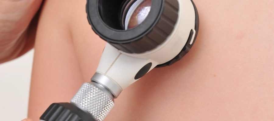

Skin cancer affects 20% of population in during the whole lifespan. Their removal is one of the most represented surgeries all over the world. In the United States, this surgery accounts for 77% of all the reconstructive plastic surgery procedures performed annually, recording more than 4.4 million operations per year.
Types of skin cancer
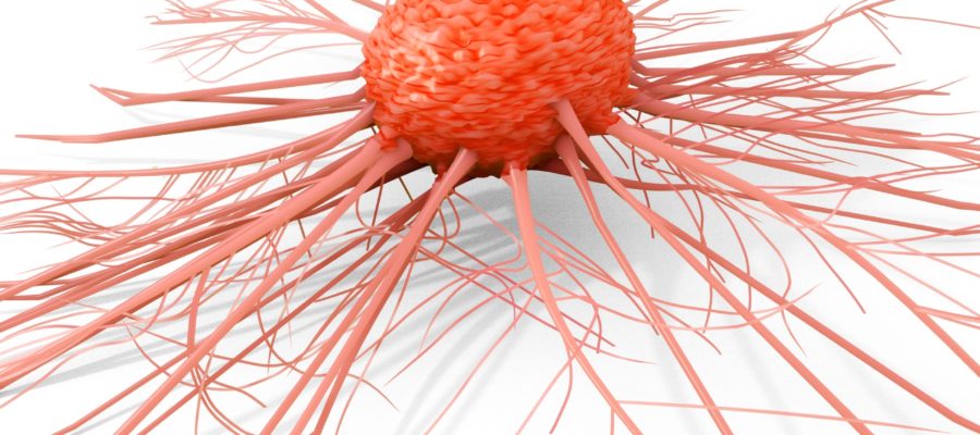
The most common skin cancer is the Basal Cell Carcinoma (BCC). This tumor represents the 80% of all skin cancers. Histologically it derives from basal keratinocytes of the epidermis.
This cancer is locally invasive because grows in the primary site without distant metastases. The main cause is a prolonged sun exposure. This explains why the incidence is higher from the fifth decade of life. In rare cases BCC may correlate to exposure to certain chemicals, syndromic forms or display a familiar pattern.
Squamous cell carcinoma (SCC) is the second most common skin cancer. Unlike basal Cell Carcinoma, could grow in the surrounding tissues. In advanced cases it can affect the lymph nodes and metastasize.
For this reason, once diagnosed, reginal lymph nodes should be investigated with Ultrasonography. In selected cases, secondary imaging (e.g. Magnetic Resonance or Computed Tomography) is required to study locoregional and distant invasion.
Melanoma is the third most common skin cancer and is treated separately in the following paragraphs.
Surgical indication
Surgery is the elective treatment of skin cancer. Basal cell carcinoma can be treated conservatively with topical chemotherapy in the following cases:
- Very small tumors
- Not aggressive type
- When the patient can not tolerate a surgical procedure due to seniority or severe comorbidities. In this case radiotherapy should be considered.
The procedure
The surgical procedure allows an en-block removal of skin cancer and the histological examination. This provides for an essential information regarding type, aggressivity and radicality of the treatment. In case of incomplete excision or reduced safety margins, a further surgical procedure is planned.
Unlike ablation treatments with liquid nitrogen and laser techniques, surgery allows a proper histological diagnosis and could assure a radical removal.
Cutaneous melanoma
This cancer is the most aggressive cutaneous malignancy and it is the third most common malignant skin tumor with an incidence of 2-3 % in the general population. This cancer is the mutation of melanocytes and it may arise from a pre-existing mole or less frequently could originate ex-novo. Risk factors are mainly a light skin phototype, multiple sunburns, personal and familial history of melanoma, the presence of multiple dysplastic nevi.
Prognosis is defined by the stage of disease. This is defined by thickness, affected lymph-nodes and distant metastases.
In the first excision the histological diagnosis is performed. In fact incisional biopsy of pigmented lesions is usually not indicated.
In a second stage, a wide resection is performed along with the biopsy of the sentinel lymph node in most cases. The margins of this excision are based on the thickness of melanoma.
The treatment protocol is customized to each case and goes beyond the aims of this discussion.
Timing of reconstruction
Reconstruction could be performed immediately after tumor removal or later, in a second surgical procedure. This issue depends on the size of the tumor, the anatomic region, the degree of invasion and the evidence of clear margins.
When these factors allow a safe excision, immediate reconstruction is performed, otherwise is preferable to wait for the histological evidence of radicality.
Reconstructive modalities
The reconstructive modality mainly depends on the size of defect and the anatomical site. This could be summarized as follows:
- Direct closure: this modality consists in the direct closure of wound margins. The resulting scar is about 2.5 times the length of the neoformation. This option is advisable when the defect is small or when skin laxity allows a wound closure without tension. In cosmetically sensitive areas other solutions should be considered to avoid profile distortion and minimize scar visibility
- Local flaps. Local flaps are composed by skin and a small portion of subcutaneous tissue. These flaps are supplied randomly by dermal vasculature and allow the reconstruction of small areas. The aesthetic outcome is good, however they require more extensive scarring than direct closure.
- Pedicled flaps. Pedicled flaps consist in a portion of tissue supplied by a pedicle (usually an artery and vein). A pedicled flap may include not only skin but also fascia and muscle. This solution allows to reconstruct medium size areas. The flap could reasonably reconstruct defects as far as the length of the pedicle.
- Free flaps. This solution allows the reconstruction of large and distant defects. They may include skin, muscle or bone segments. The pedicle of the flap is cut and re-connected (in jargon anastomosed) to the vessels of the acceptor site. Requires the use of a microscope to connect the vessels and a longer operative time.
- Skin grafts. A skin graft is a thin layer of skin that is applied over the defect. The graft is able to survive creating new vascular connections. They could be classified according their thickness in thin (0.25mm), medium-thickness (0.7 mm) and. full thickness grafts (when they include the whole cutaneous layer). Skin grafts usually require one week to take in the new site. During this time a compressive dressing is employed to allow a proper healing. In case of thin or medium size grafts, the color of the donor skin keeps a different pigmentation than the surrounding skin. Full thickness skin grafts instead are excised with a linear scar.
Postoperative period
This surgery usually requires 2-3 weeks of convalescence. Full photoprotection of the scar is advised for 9 months to avoid hyperpigmentation.
Histological examination gives a definitive diagnosis and informs whether the excision is radical.
Lasers and cold ablation do not provide the possibility of histological examination. Their application to skin tumors is therefore inappropriate.